


Your breakout healed. The redness calmed down. But the dark mark it left behind is still there, weeks later, looking like a shadow of the original problem. That mark is post-inflammatory hyperpigmentation, and it is the single most common form of hyperpigmentation that people deal with.
PIH is not the breakout. It is not scarring. It is what happens when your skin's inflammatory response leaves behind an excess deposit of melanin at the site where the inflammation occurred. The original event is over, but the pigment remains as a kind of biological record of what happened there.
Understanding how PIH forms, where it sits, and how it behaves is what separates a measured response from months of misdirected effort. It is also what prevents the most common mistake people make with it: treating it like melasma, or treating melasma like it.
What it looks like
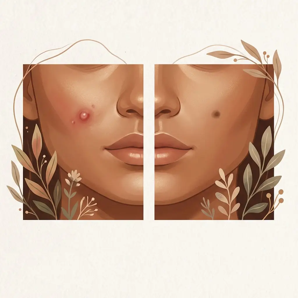
PIH appears as flat, discoloured marks at the exact location of a previous inflammatory event. The colour varies significantly depending on skin tone and where the pigment is sitting in the skin.
On lighter skin, PIH often starts as a pink or red mark that transitions to light brown over time. On medium skin tones, it tends to appear as medium brown to dark brown. On deeper skin tones, PIH can present as dark brown, deep brown, or even blue-grey when pigment has dropped into the dermal layer.
The marks are flat. If you run your finger over them, you should not feel any textural change. If there is raised tissue, an indentation, or an uneven surface, that is scarring, which is a separate issue that involves structural damage to the skin rather than excess pigment. PIH and scarring can coexist at the same site, but they are different problems.
The edges of PIH marks tend to be relatively well defined in the epidermis, because the pigment was deposited at a specific location. Deeper PIH can appear softer and more diffuse as the melanin scatters through the dermis.
Where it appears
PIH can occur anywhere on the body where inflammation has occurred. There is no region it prefers. It simply follows the trigger.
On the face, PIH is most commonly seen along the jawline, cheeks, forehead, and chin, because these are the areas most affected by acne. On the body, it can appear at sites of eczema flares, insect bites, friction injuries, burns, cuts, or any other event that generated an inflammatory response in the skin.
The location is one of the most reliable identification markers. PIH traces back to a specific event at a specific site. If you can point to where the breakout, rash, or injury was, and the dark mark sits in that exact footprint, PIH is the most likely explanation.
What triggers it
Every case of PIH starts with inflammation. The type of inflammation varies, but the mechanism is consistent: stressed or damaged skin releases inflammatory mediators, those mediators stimulate melanocytes at the site, and the melanocytes respond by producing excess melanin.
The most common triggers include:
- Acne. The most frequent cause of facial PIH, particularly inflammatory acne (papules, pustules, cysts). The worse the inflammation, the darker and more persistent the mark. Picking or squeezing adds mechanical trauma to the existing inflammation, which compounds the pigment response.
- Eczema and dermatitis flares. Any flare that produces visible redness, swelling, or irritation can leave PIH behind once it resolves. This includes atopic dermatitis, contact dermatitis, and seborrhoeic dermatitis.
- Insect bites and minor injuries. Mosquito bites, scratches, minor burns, and cuts can all produce PIH, especially in melanin-rich skin where melanocytes are more responsive to inflammatory signals.
- Cosmetic procedures. Lasers, chemical peels, microneedling, and other treatments that create controlled inflammation can trigger PIH as a side effect. This subtype is covered separately as post-procedure pigmentation because the clinical context changes the conversation.
- Skincare irritation. Over-exfoliation, acids at too-high concentrations, retinoids introduced too aggressively, or any product that creates chronic low-grade irritation. The irony of skincare causing the pigment it is trying to treat is more common than most people expect. The irritation-induced hyperpigmentation article covers this in detail.
The severity of the resulting PIH is proportional to the severity and duration of the inflammation that caused it. A mild breakout that resolves quickly may leave a faint mark that fades in weeks. A deep cystic lesion that lingers for days produces significantly more melanin and leaves a darker, more persistent mark.
How it behaves over time
PIH has a generally favourable trajectory compared to other types. Once the triggering inflammation stops, the mark is essentially a stain that the body is actively working to clear. The question is how long that process takes, and the answer varies enormously.
Mild, recent PIH in the epidermis can fade visibly within 4 to 8 weeks with no intervention at all, just through the normal cycle of skin cell turnover. With consistent topical support, that timeline can shorten.
More established or deeper PIH behaves differently. If the pigment has been there for months, or if the original inflammation was severe enough to push melanin into the dermis, the fading process slows considerably. Dermal PIH can take 6 months to 2 years to resolve, because it depends on macrophage-mediated clearance rather than simple turnover.
The important thing about PIH is that the direction is generally forward. Unlike melasma, which cycles and fluctuates, PIH that is no longer being re-triggered tends to move steadily toward resolution. Progress may be slow, but it is usually happening. If it is not, the most likely explanation is that the trigger is still active (ongoing acne, continued irritation, unprotected sun exposure recharging the melanin), the pigment is deeper than it appears, or both.
The PIH timeline covers this in detail with realistic benchmarks by severity and depth.
How deep it typically sits
Depth is what separates a PIH mark that fades in a few weeks from one that lingers for over a year, and it is often underestimated.
Epidermal PIH sits in the upper layers of the skin. The colour tends to be brown or tan with relatively defined edges. This pigment is within reach of skin turnover and topical ingredients. It responds to treatment and generally fades with time.
Dermal PIH sits deeper. The colour shifts to blue-grey, ashy, or muted brown. The edges are softer and more diffuse. At this depth, normal skin turnover cannot reach the pigment. Clearance depends on a much slower immune-mediated process where macrophages gradually remove melanin from the dermis. Topical ingredients have limited reach here, and the timeline extends to months or years.
Most PIH exists on a spectrum rather than sitting neatly in one layer. A mark can have both epidermal and dermal components, which is why the visible portion may lighten relatively quickly while a deeper shadow persists underneath.
Colour is the most accessible depth indicator. Brown and tan suggest epidermal. Blue-grey suggests dermal. On deeper skin tones, this distinction becomes harder to read visually, which is one of the reasons dermatologists sometimes use a Wood's lamp to assess pigment depth.
Relapse risk
PIH has a relatively low relapse risk compared to melasma, but "low" is not the same as zero.
Once a PIH mark has faded, it does not spontaneously return. The pigment that was deposited has been cleared, and that specific mark is resolved. This is fundamentally different from melasma, where the melanocytes remain in a state of chronic hyperactivity and can reactivate with seasonal changes, hormonal shifts, or heat exposure.
Where PIH relapse does occur, it is because the triggering condition recurs. If acne continues to flare in the same area, new marks replace old ones and the skin never gets a clear window to fully recover. If the irritating product stays in the routine, the low-grade inflammation it generates keeps producing fresh PIH on top of what is trying to heal.
The practical implication: controlling the trigger matters as much as treating the mark. A fading plan without trigger control is a plan that keeps falling behind.
How it is commonly confused
PIH vs melasma is the most consequential confusion. Both present as dark patches on the face, but PIH is event-driven, site-specific, and moves toward resolution, while melasma is hormone-driven, symmetrical, and cycles chronically. The treatment tolerance is different too. PIH generally handles more intensity. Melasma frequently rebounds under the same approach. The PIH vs Melasma comparison covers this in full.
PIH vs sun spots causes confusion when both appear as brown marks on the face. The key difference is history: PIH traces to a specific inflammatory event and fades over time. Sun spots are the result of cumulative UV exposure and remain stable indefinitely without intervention. If you can identify a triggering event, it is probably PIH. If the mark appeared gradually with no clear origin, it is more likely a sun spot.
PIH vs post-inflammatory erythema (PIE) is the confusion that wastes the most product. PIE is a red or pink mark left by inflammation, but it is vascular, not melanin-driven. It represents dilated or damaged blood vessels, not excess pigment. Brightening serums and tyrosinase inhibitors do nothing for PIE because there is no excess melanin to target. The hyperpigmentation vs redness article covers how to distinguish between the two.
PIH vs post-procedure pigmentation matters after clinical treatment. Post-procedure marks are mechanistically the same as PIH (the procedure's inflammation triggered melanocyte overproduction), but the context is different: a clinical setting, a provider's responsibility, a recovery protocol. The distinction matters because the question shifts from "what caused this?" to "was this expected, and when should I be concerned?"
When to see a dermatologist
Most PIH does not require medical intervention. It is a cosmetic concern that fades on its own or with over-the-counter support. However, there are situations where professional evaluation is worth pursuing.
If the mark has not changed at all after 6 months of consistent effort. PIH that has truly stopped progressing may have a significant dermal component that topicals cannot reach, or the initial identification may be wrong.
If new marks continue to appear and you cannot identify the trigger. Persistent new PIH without an obvious inflammatory source may point to an underlying condition (ongoing low-grade inflammation, a sensitivity you have not identified) that needs investigation.
If the original texture is also affected. Combined PIH and scarring benefits from a dermatologist's assessment of which component to address first and how to avoid worsening one while treating the other.
If you suspect it may not be PIH. Symmetrical distribution, no clear triggering event, cycling with hormones or seasons, or blue-grey colour without a history of deep inflammation are all signals that something other than straightforward PIH may be going on.
PIH is your skin's memory of inflammation. Once the inflammation stops, the memory fades. The speed depends on depth, skin tone, and whether the trigger is truly gone.


