


Lasers are the most powerful clinical tool available for hyperpigmentation. They can fragment pigment that sits deeper than any topical or peel can reach, and in the right hands on the right skin, they produce results that nothing else matches.
They also carry the highest risk profile of any pigmentation treatment. The same energy that breaks apart melanin can trigger new melanin production if the settings are wrong, the skin tone is not accounted for, or the pigment type is misidentified. Of all the clinical methods used for pigmentation, lasers are the ones most likely to worsen the condition when the approach is not well matched.
That is not a reason to avoid them. It is a reason to understand what they do, how they differ, and what determines whether the outcome is good or bad.
How Lasers Work on Pigment
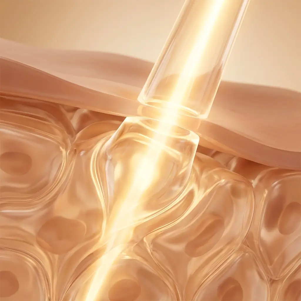
Lasers treat pigmentation through a principle called selective photothermolysis. The device emits light at a specific wavelength that is preferentially absorbed by melanin. When melanin absorbs that energy, it heats up rapidly and fragments into smaller particles. The body's immune system then clears those fragments over the following weeks.
The selectivity is the key. A well-calibrated laser targets melanin while leaving the surrounding tissue relatively undamaged. But melanin exists in all skin, not just in hyperpigmented areas. In darker skin tones, the laser has more competing melanin to interact with, which increases the risk of collateral thermal damage and the inflammatory response that follows it.
Two variables determine how a laser interacts with pigment: wavelength (which determines how deep the light penetrates and how strongly melanin absorbs it) and pulse duration (which determines how quickly the energy is delivered). Shorter pulses concentrate energy more tightly on the pigment target. Longer pulses spread the energy more diffusely, which can reduce selectivity but also reduce the risk of mechanical tissue damage.
Types of Lasers Used for Hyperpigmentation
Q-switched lasers deliver energy in nanosecond pulses (billionths of a second). They were the standard for pigment-specific laser treatment for decades and remain widely used. Common Q-switched devices include the Q-switched Nd:YAG (1064nm and 532nm), Q-switched ruby (694nm), and Q-switched alexandrite (755nm).
The 1064nm Nd:YAG wavelength is particularly relevant for darker skin tones because it penetrates deeper and is less strongly absorbed by epidermal melanin, which reduces the risk of surface damage. The 532nm wavelength targets superficial pigment more aggressively but carries higher risk in melanin-rich skin.
Picosecond lasers are the newer generation. They deliver energy in picosecond pulses (trillionths of a second), which is roughly a thousand times faster than Q-switched. The shorter pulse duration means the pigment is fragmented more by mechanical (photoacoustic) force than by heat, which in theory produces less thermal damage to surrounding tissue.
In practice, picosecond lasers have shown equivalent or slightly better results than Q-switched for many pigment conditions, with some evidence of reduced downtime and lower risk of post-inflammatory hyperpigmentation. They are not universally superior. The advantage depends on the pigment type, depth, and skin tone being treated.
Fractional lasers work differently. Rather than targeting pigment directly, they create a pattern of microscopic treatment zones (columns of damaged tissue) surrounded by untreated skin. The untreated skin accelerates healing. Fractional lasers are divided into ablative (which vaporise tissue) and non-ablative (which heat tissue without removing it).
Non-ablative fractional lasers (like fractional 1550nm or 1927nm devices) are sometimes used for pigmentation and skin rejuvenation. They can improve tone and texture gradually over a series of sessions. However, fractional lasers are generally less pigment-specific than Q-switched or picosecond devices, and for melasma they carry a high rebound risk that is covered in detail below.
Which Pigment Types and Skin Tones Respond
Discrete, well-defined spots (sun lentigines, age spots, some post-inflammatory marks) tend to respond best to laser treatment. The pigment is localised, often epidermal, and the contrast between the spot and surrounding skin gives the laser a clear target. Q-switched and picosecond lasers are typically the first choice for these.
Post-inflammatory hyperpigmentation (PIH) can respond to laser treatment, but the approach needs to be conservative. PIH indicates that the skin has already demonstrated an inflammatory pigment response. Treating it with a device that generates inflammation carries inherent risk. Low-fluence settings and longer intervals between sessions reduce that risk.
Melasma is the most challenging pigment condition to treat with lasers. Low-fluence Q-switched Nd:YAG ("laser toning") has been used for melasma with mixed results. Some patients see improvement. Others experience rebound darkening, mottled hypopigmentation, or both. The vascular component of melasma (increased blood vessel density in affected skin) adds complexity that pigment-targeting lasers alone do not address. Most specialists now consider lasers a last-resort or supporting option for melasma rather than a first-line treatment.
Skin tone is the single most important safety variable in laser treatment for pigmentation. Dermatologists use the Fitzpatrick scale (I through VI) to classify skin by its response to UV, with higher numbers indicating more melanin and greater pigment reactivity. Fitzpatrick I to III skin generally tolerates a wider range of laser types, wavelengths, and energy settings. Fitzpatrick IV to VI skin has a much narrower safety window, and the risk of post-inflammatory hyperpigmentation, hypopigmentation (permanent lightening), and scarring all increase with melanin density. The skin tone risk notes below cover the specific adjustments this requires.

Recovery and Downtime
Q-switched and picosecond lasers: Treated spots typically darken immediately after treatment (this is expected and called "frosting" or "ash white" response depending on the device and settings). Over the following 7 to 14 days, the darkened spots crust lightly and flake off, revealing lighter skin underneath. There may be mild redness and sensitivity during this period. Most people return to normal activities within a day or two, though the treated areas remain visible during the shedding phase.
Fractional lasers: Recovery is more involved. Expect redness, swelling, and a rough or sandpaper-like texture for 3 to 7 days (non-ablative) or 7 to 14 days (ablative). The skin may ooze lightly in the first day or two with ablative fractional treatment. Makeup is typically avoided for at least a few days.
Across all laser types, the skin is much more vulnerable to UV damage during the healing period. Sun exposure during recovery is one of the most common causes of post-treatment hyperpigmentation. Rigorous sun protection is essential for the entire healing window and for several weeks after.
Risk Profile
Who should be cautious or avoid this (for now):
- Anyone with an active tan or recent significant sun exposure (increases melanin density in the epidermis, raising risk of burns and PIH)
- Anyone currently using photosensitising medications (certain antibiotics, retinoids, some anti-inflammatories)
- Anyone with active skin infections or a damaged barrier in the treatment area
- Anyone with a history of keloid scarring (ablative lasers carry the highest risk)
- Anyone with melasma who has not tried conservative options first (topicals, sun protection, trigger management)
Skin tone risk notes:
Laser treatment carries the highest skin-tone-dependent risk of any pigmentation procedure. Most laser devices were developed and calibrated on Fitzpatrick I to III skin. Using them on darker skin without adjusting wavelength, fluence, and pulse duration accordingly can cause burns, PIH, or permanent hypopigmentation. The 1064nm Nd:YAG wavelength is the safest option for Fitzpatrick IV to VI skin. Even with appropriate settings, the margin for error is narrower, and practitioner experience with diverse skin tones is not negotiable.
Rebound risk:
Rebound pigmentation after laser treatment is well documented, particularly for melasma and hormonally influenced pigmentation. The laser fragments existing pigment, but if the underlying triggers (hormonal, inflammatory, UV-related) remain active, melanocytes will produce new pigment to replace what was removed. Post-treatment PIH can also occur independently: the laser-induced inflammation itself triggers new melanin production in susceptible skin. This typically appears 2 to 6 weeks after treatment and can be darker than the original pigmentation.
Questions to ask your provider:
- Which laser device and wavelength do you recommend for my specific pigment type and skin tone?
- What is your experience treating Fitzpatrick IV to VI skin with this device?
- What energy settings will you use, and how do you adjust for melanin-rich skin?
- What is the realistic risk of post-inflammatory hyperpigmentation or hypopigmentation with this protocol?
- How many sessions do you typically recommend, and what intervals do you use between them?
- What happens if the pigment comes back or worsens after treatment?
Best paired with:
Laser treatment produces the best outcomes when the skin is well prepared and well supported afterwards. A consistent protection and prevention routine is essential before, during, and after treatment. Many practitioners also prescribe pre-treatment topicals (hydroquinone, retinoids, or both) for 4 to 6 weeks before laser to reduce melanocyte activity and lower the risk of post-treatment rebound. Post-treatment, topical brightening actives (vitamin C, niacinamide, tranexamic acid) are typically resumed once the skin has healed.
Pre-treatment topicals prepare the skin at the surface. What they cannot prepare is the internal environment the melanocytes are sitting in. Laser treatment delivers concentrated energy that the body must process through its inflammatory and repair pathways. If systemic inflammation is already elevated, if oxidative stress is high, or if the micronutrients required for cellular repair are depleted, the skin's response to that energy skews toward rebound rather than resolution. This is part of why identical laser settings produce different outcomes on different people with similar skin tones. The surface was matched, but the internal conditions were not. The signalling-layer pages cover what drives melanocyte behaviour beneath the epidermis.

The Takeaway
Lasers can reach pigment that nothing else can. For the right type of hyperpigmentation on the right skin, they produce results that topicals, peels, and supplements cannot match. That's their genuine strength.
But they are also the treatment where the gap between a good outcome and a bad one is widest. The device matters. The wavelength matters. The settings matter. The practitioner's experience with your specific skin tone and pigment type matters more than all of those combined. The right consultation is worth more than the treatment itself.


