


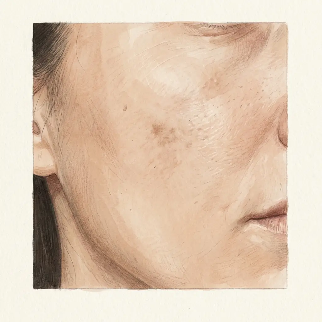
The breakout cleared. The bump is gone. But in its place there is a dark mark that was not there before, and it looks like it has no intention of leaving.
This is post-inflammatory hyperpigmentation from acne. It is the most common form of PIH, the most predictable, and for many people the more lasting problem than the acne itself. The breakout is temporary. The mark it leaves behind can persist for months, sometimes longer, depending on how deep the inflammation went and what the skin did in response.
Understanding the mechanism does not just explain why the mark is there. It explains why some breakouts leave marks and others do not, why the ones near your jawline are darker than the ones on your forehead, and why every time you picked at one it came back worse.
What happens inside a breakout
A breakout is an inflammatory event. Even a small whitehead involves a localised immune response: bacteria multiply inside a blocked pore, the immune system detects the threat, and inflammatory mediators flood the area to contain it. Neutrophils arrive. Cytokines accumulate. The tissue swells. That is the bump you see and feel.
Melanocytes are bystanders in this process. They are not part of the immune response. But they sit in the epidermis directly adjacent to where the inflammation is happening, and they are sensitive to the same inflammatory signals the immune system is producing.
Prostaglandins, leukotrienes, and interleukins released during the breakout do not stay neatly contained within the pore. They diffuse into the surrounding tissue. When they reach the melanocytes nearby, those melanocytes interpret the inflammatory signals as a reason to produce melanin. The biological logic is protective: melanin has antioxidant and free-radical-scavenging properties, so the body produces it in areas of tissue stress. But the practical result is a dark mark at the site of every lesion where the inflammation was strong enough to reach the melanocytes.
The breakout does not need to be severe for this to happen. It just needs to be inflammatory enough.
That threshold is lower than most people expect.
Why depth and severity determine the mark
Not every breakout leaves a dark mark. The variable is how much inflammatory signalling reaches the melanocytes, and that depends primarily on two things: how deep the lesion sits and how intense the immune response is.
A superficial whitehead that forms near the skin surface, resolves in a few days, and produces minimal swelling generates a limited inflammatory radius. The signals may not reach the melanocytes in sufficient concentration to trigger a visible pigment response. It comes and goes without leaving a trace.
A deep cystic lesion is a different event entirely. The inflammation originates deeper in the dermis, involves a larger volume of tissue, and persists for longer. The inflammatory mediators are produced in higher concentrations and diffuse over a wider area. More melanocytes are exposed. They are exposed for longer. And the pigment response is proportionally larger.
This is why the deep, painful breakouts along the jawline and lower cheeks tend to leave the most persistent marks. They are not just more visible. They are producing more melanin, at greater depth, in response to a more intense and prolonged inflammatory response. The mark is proportional to the event that caused it.
Nodular acne takes this further. When the follicle wall ruptures beneath the surface and the contents spill into surrounding tissue, the immune response escalates sharply. The inflammation is no longer contained within a pore. It is a tissue-level event. The pigment response is correspondingly more intense and more likely to involve dermal melanin deposition, which is below the reach of normal epidermal turnover and takes much longer to clear.
Why picking and squeezing makes it worse
You already know you are not supposed to pick. This is why it matters for pigmentation specifically.
When you squeeze or pick at a breakout, you are applying mechanical force to tissue that is already inflamed. That force does three things that each independently worsen the pigment outcome.
First, it ruptures the follicle wall. A lesion that might have resolved within its contained space now leaks its inflammatory contents, bacteria, sebum, dead cells, into surrounding tissue. The immune response escalates because the threat is no longer contained. More inflammatory mediators are produced. More melanocytes are reached.
Second, it damages tissue at the surface. The squeezing creates micro-tears in the epidermis and dermis. Tissue damage triggers its own inflammatory response independent of the acne itself. The melanocytes now receive inflammatory signals from two sources: the breakout underneath and the mechanical injury on top. The pigment response reflects the total inflammatory load, not just the original lesion.
Third, it disrupts the local barrier. The compromised skin at the picking site is more permeable to UV and more prone to further irritation during the healing window. Every additional insult during that period adds to the cumulative inflammatory signalling that melanocytes are responding to. Aggressive topical acne treatments applied to actively inflamed, picked skin can produce a similar compounding effect, which why treatments sometimes backfire covers in detail.
The mark left by a picked breakout is almost always darker, larger, and longer-lasting than the mark the same breakout would have left on its own. The additional pigment is not from the acne. It is from the intervention.
The breakout was one event. The picking made it two.
Why some skin marks more than others
Two people can have the same breakout pattern and leave completely different marks. One clears without a trace. The other is left with dark spots at every site. The breakouts were similar. The pigment response was not.
Skin tone is the most visible factor. In darker skin tones (Fitzpatrick IV to VI), melanocytes are more numerous, more active at baseline, and more responsive to inflammatory signalling. The same inflammatory stimulus produces a proportionally larger melanin output. This is why acne-related PIH is especially common and persistent in melanin-rich skin. The acne may be identical. The pigment aftermath is not.
But skin tone is not the only variable. People with lighter skin tones who mark heavily and people with darker skin tones who rarely mark both exist. The difference is partly in the inflammatory environment the melanocytes are operating in at the time of the breakout.
When background systemic inflammation is elevated, whether from stress, poor sleep, diet, hormonal fluctuation, or other inflammatory conditions, the melanocytes are already in a heightened state of reactivity before the breakout even forms. The inflammatory signals from the breakout arrive in a system that is already primed. The threshold for triggering a pigment response is lower. Breakouts that would have resolved without a mark in a low-inflammation state now leave visible pigmentation because the cumulative inflammatory load crossed the activation threshold.
This is why acne-related PIH often worsens during stressful periods, hormonal shifts, or when other inflammatory conditions are flaring. The breakouts may not be worse. The pigment response is, because the systemic context has changed.
If your breakouts have started leaving marks when they did not used to, and the acne itself has not changed, something in the background may have. Hyperpigmentation from within covers what those background conditions are and what influences them.
What influences whether marks fade or persist
Once the breakout resolves, the mark is there. What happens next depends on where the pigment sits and how effectively the body clears it.
Epidermal pigment, deposited in the upper layers, migrates upward with normal cell turnover and sheds at the surface over roughly four to eight weeks. This is the pigment that responds to topical brightening treatment. Most mild acne PIH sits here.
Dermal pigment, dropped below the basement membrane during intense inflammation, is cleared by macrophages through a slow phagocytic process. It does not respond to surface turnover. It can take months to years to clear, and topical treatment has limited reach. This is the pigment that makes some marks feel permanent even though they are technically still fading.
The speed of the inflammatory resolution after the breakout determines how much pigment ends up in each layer. Inflammation that resolves quickly limits the total melanin produced and keeps most of it epidermal. Inflammation that smoulders for weeks produces more melanin over a longer period and increases the chance of dermal deposition.
That resolution speed depends on the same systemic variables that influenced the initial response: anti-inflammatory substrate availability, oxidative stress load, and the capacity of the body's pro-resolving pathways to switch the immune response from active inflammation to tissue repair. When those resources are depleted or insufficient, each breakout's aftermath stretches longer, produces more pigment, and the marks accumulate faster than they fade.
Kallistia's internal support is formulated around these resolution and regulation variables: the anti-inflammatory capacity, antioxidant availability, and pro-resolving substrates that influence both how strongly melanocytes respond to each breakout and how quickly the inflammatory aftermath settles. For acne-related PIH, the internal layer does not treat the acne or fade the mark directly. It addresses the systemic conditions that determine how much pigment each breakout produces and how long it stays.
The takeaway
Acne triggers pigmentation through the same inflammatory response that makes the breakout visible in the first place. The melanocytes are bystanders that respond to inflammatory signals by producing melanin at the lesion site. The deeper and more intense the inflammation, the more pigment is produced and the deeper it is deposited.
Picking compounds the response by adding mechanical inflammation on top of the acne inflammation. The mark from a picked breakout reflects both events.
How much pigment each breakout ultimately produces, and how long it persists, depends on two things: the severity of the inflammatory event itself, and the systemic inflammatory environment that determines how reactive the melanocytes were at the time and how quickly the aftermath resolves. The first is the breakout. The second is everything else.


